images that haunt us
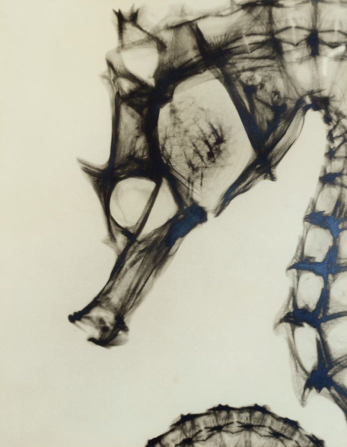
Anonymous maker. Radiographie d’hippocampe / X-ray of a Sea Horse, around 1930
/ src
lumieredesroses
more [+] sea horses


All images retrieved from: A system of instruction in X-ray methods and medical uses of light, hot-air, vibration and high-frequency currents : a pictorial system of teaching by clinical instruction plates with explanatory text : a series of photographic clinics in standard uses of scientific therapeutic apparatus for surgical and medical practitioners : prepared especially for the post-graduate home study of surgeons, general physicians, dentists, dermatologists and specialists in the treatment of chronic diseases, and sanitarium practice (New York, 1902) by S. H. (Samuel Howard) Monell
Topics: Vibration, X-rays, Diagnosis, Radioscopic, Thermotherapy, Electrotherapeutics, X-Ray Therapy, Vibration, Diagnosis
source internet archive (Harvard Medical Library)


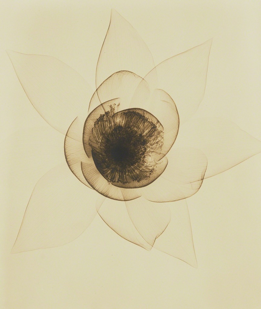
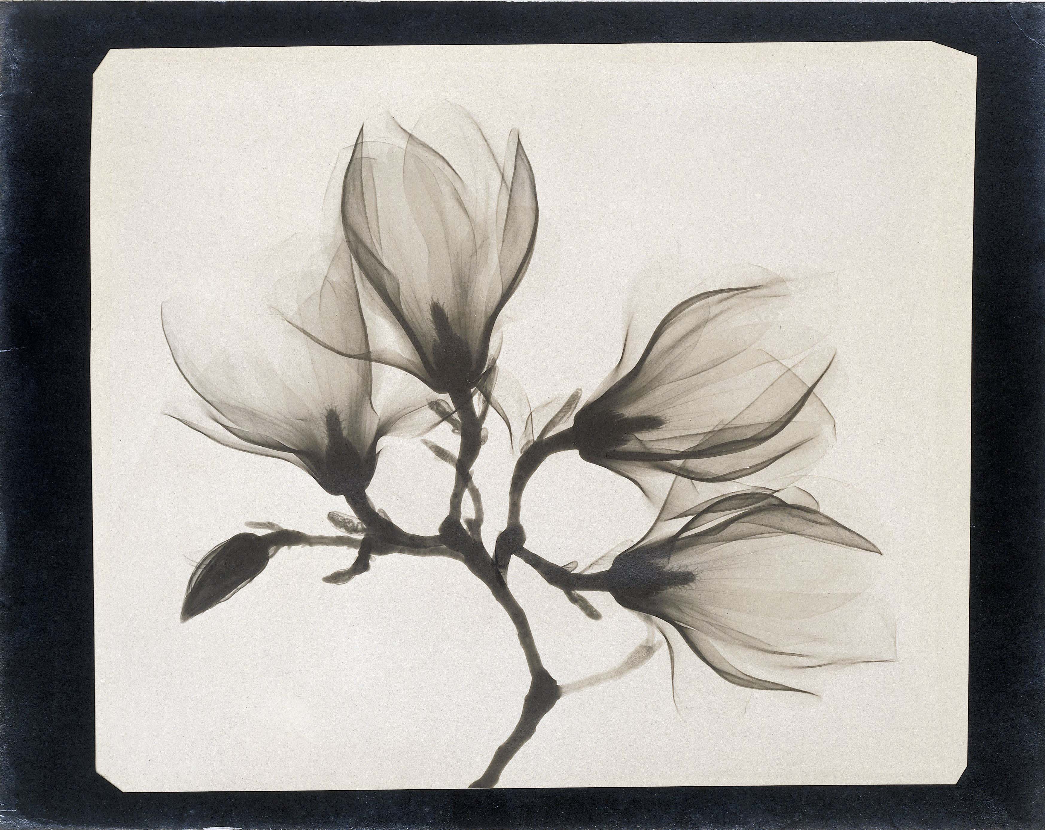
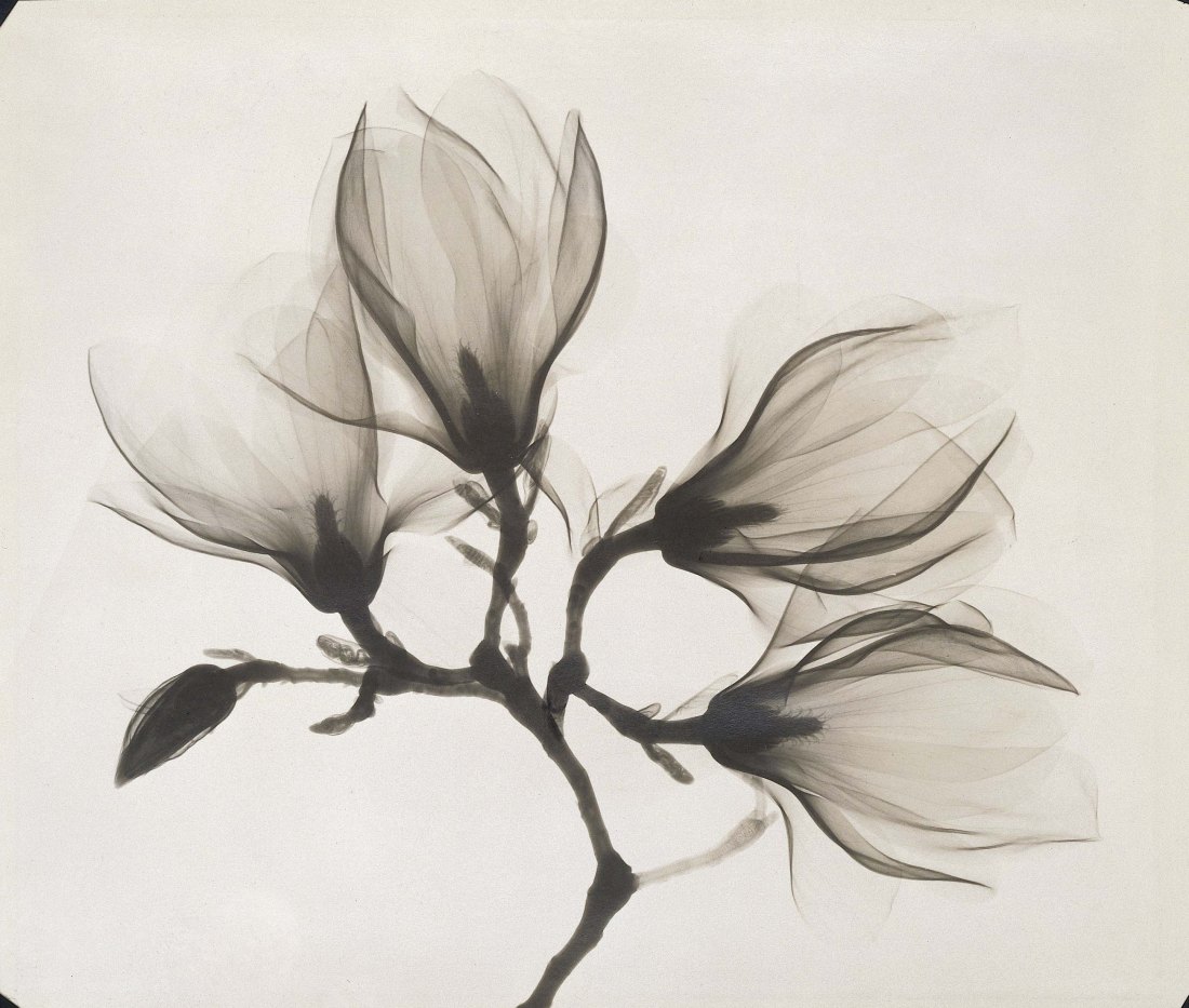
Dain Tasker :: Amazon-Lily, circa 1930. Silver print from an x-ray negative. / via
more [+] by this photographer
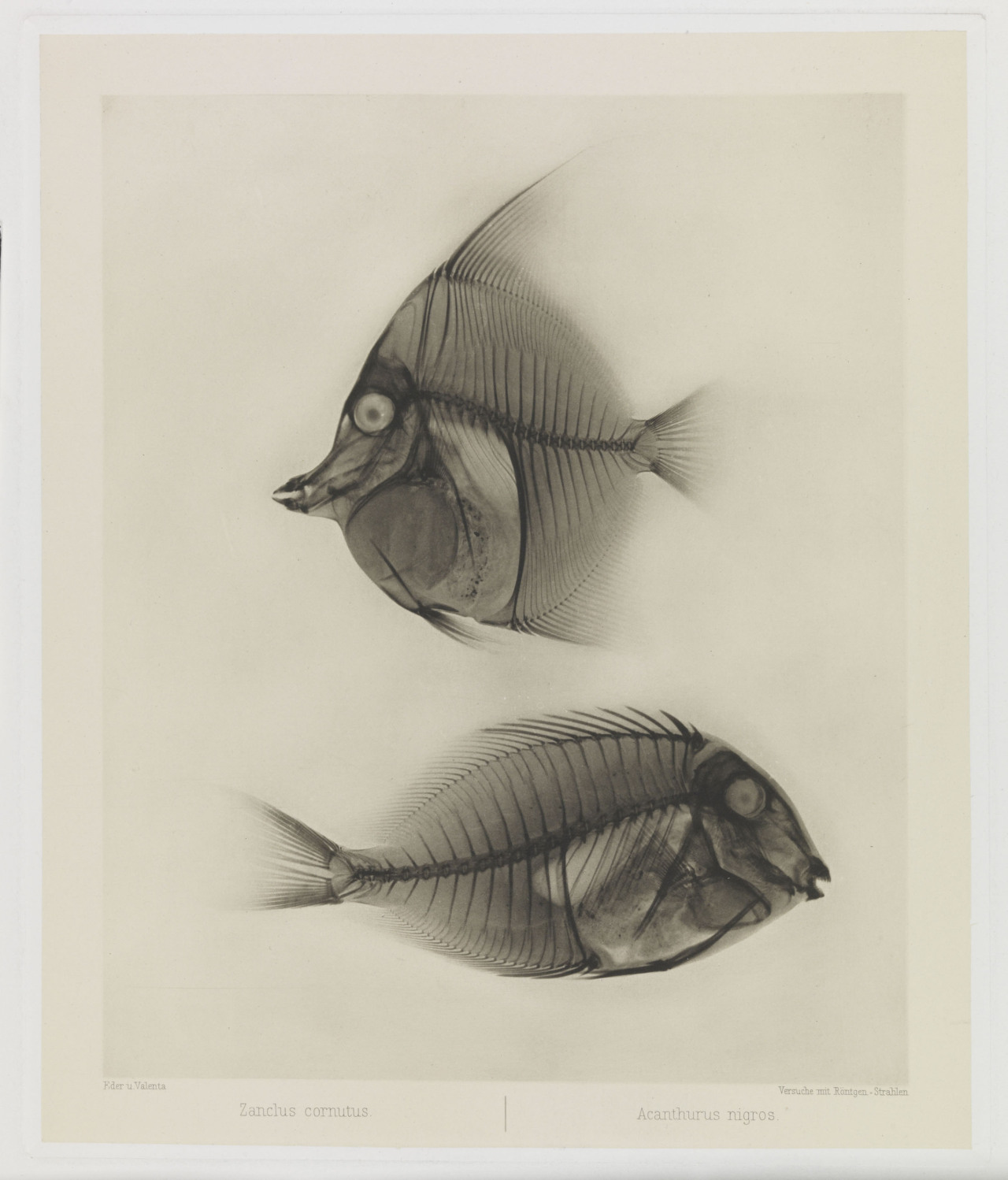
Eduard Valenta & Josef Maria Eder :: X Ray of Angelfish and Surgeonfish (Zanclus cornutus and Acanthurus nigros), 1896 [Photogravure] / src: The Met
“Eder was the director of an institute for graphic processes and the author of an early history of photography. With the photochemist Valenta, he produced a portfolio in January 1896, less than a month after Wilhelm Conrad Röntgen published his discovery of X-rays. Eder and Valenta’s volume, from which this plate derives, demonstrated the X-ray’s magical ability to reveal the hidden structure of living things.” src: The Met

Dain Tasker :: Lily, 1930 (vintage gelatin silver print) / related post, here
source: GittermanGallery